Please log in to purchase this product.
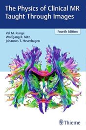
The Physics of Clinical MR Taught Through Images, 4ed (PDF)
Please log in to view the price.
The Physics of Clinical MR Taught Through Images, 4ed (PDF)
The Physics of Clinical MR Taught Through Images Fourth Edition by Val Runge, Wolfgang Nitz, and Johannes Heverhagen presents a unique and highly practical approach to understanding the physics of magnetic resonance imaging. Each physics topic is described in user-friendly language and accompanied by high-quality graphics and/or images. The visually rich format provides a readily accessible tool for learning, leveraging, and mastering the powerful diagnostic capabilities of MRI.
Key Features
- More than 700 images, anatomical drawings, clinical tables, charts, and diagrams, including magnetization curves and pulse sequencing, facilitate acquisition of highly technical content.
- Eight systematically organized sections cover core topics: hardware and radiologic safety; basic image physics; basic and advanced image acquisition; flow effects; techniques specific to the brain, heart, liver, breast, and cartilage; management and reduction of artifacts; and improvements in MRI diagnostics and technologies.
- Cutting-edge topics including contrast-enhanced MR angiography, spectroscopy, perfusion, and advanced parallel imaging/data sparsity techniques.
- Discussion of groundbreaking hardware and software innovations, such as MR-PET, 7 T, interventional MR, 4D flow, CAIPIRINHA, radial acquisition, simultaneous multislice, and compressed sensing.
- A handy appendix provides a quick reference of acronyms, which often differ from company to company.
The breadth of coverage, rich visuals, and succinct text make this manual the perfect reference for radiology residents, practicing radiologists, researchers in MR, and technologists.
Related Products
Radiology Books
Targeted Cancer Imaging: Design and Synthesis of Nanoplatforms based on Tumor Biology (EPUB)
Radiology Books
Neuroimaging in Parkinson’s Disease and Related Disorders (Original PDF from Publisher)
Radiology Books
Radiology Books
Radiology Books
American journal of Neuroradiology 2023 Full Archives (True PDF)
Radiology Books
Radiology Books
Radiology Books
JFR Plus 2023 (JOURNÉES FRANCOPHONES DE RADIOLOGIE DIAGNOSTIQUE & INTERVENTIONNELLE) (Videos)
Radiology Books
JFR Plus 2022 (JOURNÉES FRANCOPHONES DE RADIOLOGIE DIAGNOSTIQUE & INTERVENTIONNELLE) (Videos)
Radiology Books
American Journal of Roentogelogy 2023 Full Archives (True PDF)
Radiology Books
Contemporary Diagnostic Radiology 2023 Full Archives (True PDF)
Radiology Books
Textbook of Radiology and Imaging, 2 Volume Set, 8th edition (azw3+ePub+Converted PDF)
Radiology Books
Radiology Books
Medical Image Analysis (The MICCAI Society book Series) (Original PDF from Publisher)
Radiology Books
Advances in Medical Imaging, Detection, and Diagnosis (EPUB)
Radiology Books
Radiology Neuroradiology: Board and Certification Review, 7th Edition (AZW3 + EPUB + Converted PDF)
ORTHOPAEDICS SURGERY
PLASTIC & RECONSTRUCTIVE SURGERY
The Aesthetic Society Nuances in Injectables The Next Beauty Frontier 2022
Radiology Books
Radiology Books
Critical Case Findings and Practice Management in the ED Online Course 2022 (CME VIDEOS)
Radiology Books
Radiology Books
Challenging Cases in Thoracic Imaging: Unknown Film Panel Session Online Course 2022 (CME VIDEOS)
Radiology Books
Radiology Books
Reading Cases with the Experts: An Interactive Session Online Course 2022 (CME VIDEOS)
Radiology Books
Tumor Imaging: CT Colonography Online Course 2022 (CME VIDEOS)
Radiology Books
Radiology Books
Radiology Books
Radiology Books
Radiology Books
Multimodality Imaging, Volume 1 (Original PDF from Publisher)
Radiology Books
Frontiers of Artificial Intelligence in Medical Imaging (Original PDF from Publisher)
Radiology Books
Radiology Books
Absolute Breast Imaging Review (Original PDF from Publisher)
Radiology Books
Fuzzy Sets Methods in Image Processing and Understanding (Original PDF from Publisher)
Radiology Books
Radiology Books
American journal of Neuroradiology 2022 Full Archives (True PDF)
Radiology Books
How to Read a Normal Scan : Brain CT (High Quality Image PDF)
Radiology Books
How to read a Normal Scan: Spine MRI (High Quality Image PDF)
Radiology Books
How to Read a Normal Scan : SPINE CT (High Quality Image PDF)
Radiology Books
Magnetic Resonance Imaging of The Pelvis: A Practical Approach (Original PDF from Publisher)
Radiology Books
Radiology Books
Fuzzy Sets Methods in Image Processing and Understanding (EPUB)
Radiology Books
Radiology Books
Radiology Books
Radiology Books
Ultrasound Guided Vascular Access: Practical Solutions to Bedside Clinical Challenges (EPUB)
Radiology Books
Radiology Books
Workbook for Textbook of Diagnostic Sonography, 9th Edition (Original PDF from Publisher)
Radiology Books
Radiology Books
Radiology Books
How to Do An Echo Exam: Third Edition (Echocardiography Illustrated) (Original PDF from Publisher)
Radiology Books
Imaging for Students, 5th Edition (Original PDF from Publisher)
Radiology Books
Radiology Books
Radiology Books
Eco-Doppler Vascular: Guia Prático (Original PDF from Publisher)
Radiology Books
Contemporary Diagnostic Radiology 2021 Full Archives (True PDF)
Radiology Books
Radiology Books
Radiology Books
American Journal of Roentogelogy 2022 Full Archives (True PDF)
Radiology Books
Radiology Books
Radiology Books
Radiology Books
Radiology Books
Radiology Books
Radiology Books
Radiology Books
Radiology Books
Top 3 Differentials in Pediatric Radiology: A Case Review (EPUB)
Radiology Books
Top 3 Differentials in Nuclear Medicine: A Case Review (EPUB)

