Please log in to purchase this product.
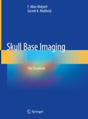
Skull Base Imaging The Essentials 2020 ORIGINAL PDF
Please log in to view the price.
Skull Base Imaging The Essentials 2020 ORIGINAL PDF
This book is a comprehensive guide to skull base imaging. Skull base is often a “no man’s land” that requires treatment using a team approach between neurosurgeons, head and neck surgeons, vascular interventionalists, radiotherapists, chemotherapists, and other professionals. Imaging of the skull base can be challenging because of its intricate anatomy and the broad breadth of presenting pathology. Although considerably complex, the anatomy is comparatively constant, while presenting pathologic entities may be encountered at myriad stages. Many of the pathologic processes that involve the skull base are rare, causing the average clinician to require help with their diagnosis and treatment. But, before any treatment can begin, these patients must come to imaging and receive the best test to establish the correct diagnosis and make important decisions regarding management and treatment. This book provides a guide to neuoradiologists performing that imaging and as a reference for related physicians and surgeons.
The book is divided into nine sections: Pituitary Region, Cerebellopontine Angle, Anterior Cranial Fossa, Middle Cranial Fossa, Craniovertebral Junction, Posterior Cranial Fossa, Inflammatory, Sarcomas, and Anatomy. Within each section, either common findings in those skull areas or different types of sarcomas or inflammatory conditions and their imaging are detailed. The anatomy section gives examples of normal anatomy from which to compare findings against. All current imaging techniques are covered, including: CT, MRI, US, angiography, CT cisternography, nuclear medicine and plain film radiography. Each chapter additionally includes key points, classic clues, incidence, differential diagnosis, recommended treatment, and prognosis.
Skull Base Imaging provides a clear and concise reference for all physicians who encounter patients with these complex and relatively rare maladies.
Related Products
Radiology Books
Targeted Cancer Imaging: Design and Synthesis of Nanoplatforms based on Tumor Biology (EPUB)
Radiology Books
Neuroimaging in Parkinson’s Disease and Related Disorders (Original PDF from Publisher)
Radiology Books
Radiology Books
Radiology Books
American journal of Neuroradiology 2023 Full Archives (True PDF)
Radiology Books
Radiology Books
Radiology Books
JFR Plus 2023 (JOURNÉES FRANCOPHONES DE RADIOLOGIE DIAGNOSTIQUE & INTERVENTIONNELLE) (Videos)
Radiology Books
JFR Plus 2022 (JOURNÉES FRANCOPHONES DE RADIOLOGIE DIAGNOSTIQUE & INTERVENTIONNELLE) (Videos)
Radiology Books
American Journal of Roentogelogy 2023 Full Archives (True PDF)
Radiology Books
Contemporary Diagnostic Radiology 2023 Full Archives (True PDF)
Radiology Books
Textbook of Radiology and Imaging, 2 Volume Set, 8th edition (azw3+ePub+Converted PDF)
Radiology Books
Radiology Books
Medical Image Analysis (The MICCAI Society book Series) (Original PDF from Publisher)
Radiology Books
Advances in Medical Imaging, Detection, and Diagnosis (EPUB)
Radiology Books
Radiology Neuroradiology: Board and Certification Review, 7th Edition (AZW3 + EPUB + Converted PDF)
ORTHOPAEDICS SURGERY
PLASTIC & RECONSTRUCTIVE SURGERY
The Aesthetic Society Nuances in Injectables The Next Beauty Frontier 2022
Radiology Books
Radiology Books
Critical Case Findings and Practice Management in the ED Online Course 2022 (CME VIDEOS)
Radiology Books
Radiology Books
Challenging Cases in Thoracic Imaging: Unknown Film Panel Session Online Course 2022 (CME VIDEOS)
Radiology Books
Radiology Books
Reading Cases with the Experts: An Interactive Session Online Course 2022 (CME VIDEOS)
Radiology Books
Tumor Imaging: CT Colonography Online Course 2022 (CME VIDEOS)
Radiology Books
Radiology Books
Radiology Books
Radiology Books
Radiology Books
Multimodality Imaging, Volume 1 (Original PDF from Publisher)
Radiology Books
Frontiers of Artificial Intelligence in Medical Imaging (Original PDF from Publisher)
Radiology Books
Radiology Books
Absolute Breast Imaging Review (Original PDF from Publisher)
Radiology Books
Fuzzy Sets Methods in Image Processing and Understanding (Original PDF from Publisher)
Radiology Books
Radiology Books
American journal of Neuroradiology 2022 Full Archives (True PDF)
Radiology Books
How to Read a Normal Scan : Brain CT (High Quality Image PDF)
Radiology Books
How to read a Normal Scan: Spine MRI (High Quality Image PDF)
Radiology Books
How to Read a Normal Scan : SPINE CT (High Quality Image PDF)
Radiology Books
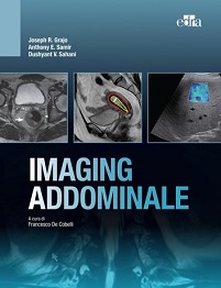
Magnetic Resonance Imaging of The Pelvis: A Practical Approach (Original PDF from Publisher)
Radiology Books
Radiology Books
Fuzzy Sets Methods in Image Processing and Understanding (EPUB)
Radiology Books
Radiology Books
Radiology Books
Radiology Books
Ultrasound Guided Vascular Access: Practical Solutions to Bedside Clinical Challenges (EPUB)
Radiology Books
Radiology Books
Workbook for Textbook of Diagnostic Sonography, 9th Edition (Original PDF from Publisher)
Radiology Books
Radiology Books
Radiology Books
How to Do An Echo Exam: Third Edition (Echocardiography Illustrated) (Original PDF from Publisher)
Radiology Books
Imaging for Students, 5th Edition (Original PDF from Publisher)
Radiology Books
Radiology Books
Radiology Books
Eco-Doppler Vascular: Guia Prático (Original PDF from Publisher)
Radiology Books
Contemporary Diagnostic Radiology 2021 Full Archives (True PDF)
Radiology Books
Radiology Books
Radiology Books
American Journal of Roentogelogy 2022 Full Archives (True PDF)
Radiology Books
Radiology Books
Radiology Books
Radiology Books
Radiology Books
Radiology Books
Radiology Books
Radiology Books
Radiology Books
Top 3 Differentials in Pediatric Radiology: A Case Review (EPUB)
Radiology Books
Top 3 Differentials in Nuclear Medicine: A Case Review (EPUB)

