Please log in to purchase this product.
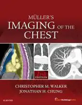
Muller’s Imaging of the Chest E-Book : Expert Radiology Series (Original PDF) 2018
Please log in to view the price.
Muller’s Imaging of the Chest E-Book : Expert Radiology Series (Original PDF) 2018
Reflecting recent major advances in the field, Müller’s Imaging of the Chest, 2nd Edition, remains your go-to reference for all aspects of chest radiology , including the latest diagnostic modalities and interventional techniques. This exhaustive resource begins with a review of normal anatomy, progressing to expert coverage based first on how patients present in clinical practice, then on diagnosis or diagnostic category. This practical, easy-to-use format helps you effectively select and interpret the best imaging studies for the everyday challenges you face in thoracic imaging. Provides extensive new information on lung cancer screening , detailing the technique required to perform a lung cancer screening CT as well as how to interpret these examinations using ACR Lung-RADS. Contains four all-new chapters : Idiopathic pleuroparenchymal fibroelastosis, Interstitial pneumonia with autoimmune features, Non-infectious complications of lung and stem cell transplantation, and Leukemia. Updates you on recent advances regarding interstitial lung disease diagnosis, diffuse idiopathic pulmonary neuroendocrine cell hyperplasia (DIPNECH), interstitial pneumonia with autoimmune features (IPAF), pleuroparenchymal fibroelastosis, and much more. Explains the recent CT classification in usual interstitial pneumonia/idiopathic pulmonary fibrosis (UIP/IPF) diagnosis and what features are required to correctly categorize a CT into one of four specific patterns. Covers current topics such as bacterial, viral, fungal, and parasitic infections, and new staging and histologic classifications for various lung neoplasms including lung cancer and mesothelioma. Features more than 3,100 superior, large digital-quality images (many in full color) depicting all of the chest imaging findings you’re likely to see, and helping you distinguish between conditions with similar manifestations. Provides boxes highlighting key points to assist you with report writing, as well as suggestions for treatment and future imaging studies. Features a full-color design throughout , color-coded tables, classic signs boxes, and bulleted lists that highlight key concepts and get you to the information you need quickly. Enhanced eBook version included with purchase. Your enhanced eBook allows you to access all of the text, figures, and references from the book on a variety of devices.
Related Products
Radiology Books
Targeted Cancer Imaging: Design and Synthesis of Nanoplatforms based on Tumor Biology (EPUB)
Radiology Books
Neuroimaging in Parkinson’s Disease and Related Disorders (Original PDF from Publisher)
Radiology Books
Radiology Books
Radiology Books
American journal of Neuroradiology 2023 Full Archives (True PDF)
Radiology Books
Radiology Books
Radiology Books
JFR Plus 2023 (JOURNÉES FRANCOPHONES DE RADIOLOGIE DIAGNOSTIQUE & INTERVENTIONNELLE) (Videos)
Radiology Books
JFR Plus 2022 (JOURNÉES FRANCOPHONES DE RADIOLOGIE DIAGNOSTIQUE & INTERVENTIONNELLE) (Videos)
Radiology Books
American Journal of Roentogelogy 2023 Full Archives (True PDF)
Radiology Books
Contemporary Diagnostic Radiology 2023 Full Archives (True PDF)
Radiology Books
Textbook of Radiology and Imaging, 2 Volume Set, 8th edition (azw3+ePub+Converted PDF)
Radiology Books
Radiology Books
Medical Image Analysis (The MICCAI Society book Series) (Original PDF from Publisher)
Radiology Books
Advances in Medical Imaging, Detection, and Diagnosis (EPUB)
Radiology Books
Radiology Neuroradiology: Board and Certification Review, 7th Edition (AZW3 + EPUB + Converted PDF)
ORTHOPAEDICS SURGERY
PLASTIC & RECONSTRUCTIVE SURGERY
The Aesthetic Society Nuances in Injectables The Next Beauty Frontier 2022
Radiology Books
Radiology Books
Critical Case Findings and Practice Management in the ED Online Course 2022 (CME VIDEOS)
Radiology Books
Radiology Books
Challenging Cases in Thoracic Imaging: Unknown Film Panel Session Online Course 2022 (CME VIDEOS)
Radiology Books
Radiology Books
Reading Cases with the Experts: An Interactive Session Online Course 2022 (CME VIDEOS)
Radiology Books
Tumor Imaging: CT Colonography Online Course 2022 (CME VIDEOS)
Radiology Books
Radiology Books
Radiology Books
Radiology Books
Radiology Books
Multimodality Imaging, Volume 1 (Original PDF from Publisher)
Radiology Books
Frontiers of Artificial Intelligence in Medical Imaging (Original PDF from Publisher)
Radiology Books
Radiology Books
Absolute Breast Imaging Review (Original PDF from Publisher)
Radiology Books
Fuzzy Sets Methods in Image Processing and Understanding (Original PDF from Publisher)
Radiology Books
Radiology Books
American journal of Neuroradiology 2022 Full Archives (True PDF)
Radiology Books
How to Read a Normal Scan : Brain CT (High Quality Image PDF)
Radiology Books
How to read a Normal Scan: Spine MRI (High Quality Image PDF)
Radiology Books
How to Read a Normal Scan : SPINE CT (High Quality Image PDF)
Radiology Books
Magnetic Resonance Imaging of The Pelvis: A Practical Approach (Original PDF from Publisher)
Radiology Books
Radiology Books
Fuzzy Sets Methods in Image Processing and Understanding (EPUB)
Radiology Books
Radiology Books
Radiology Books
Radiology Books
Ultrasound Guided Vascular Access: Practical Solutions to Bedside Clinical Challenges (EPUB)
Radiology Books
Radiology Books
Workbook for Textbook of Diagnostic Sonography, 9th Edition (Original PDF from Publisher)
Radiology Books
Radiology Books
Radiology Books
How to Do An Echo Exam: Third Edition (Echocardiography Illustrated) (Original PDF from Publisher)
Radiology Books
Imaging for Students, 5th Edition (Original PDF from Publisher)
Radiology Books
Radiology Books
Radiology Books
Eco-Doppler Vascular: Guia Prático (Original PDF from Publisher)
Radiology Books
Contemporary Diagnostic Radiology 2021 Full Archives (True PDF)
Radiology Books
Radiology Books
Radiology Books
American Journal of Roentogelogy 2022 Full Archives (True PDF)
Radiology Books
Radiology Books
Radiology Books
Radiology Books
Radiology Books
Radiology Books
Radiology Books
Radiology Books
Radiology Books
Top 3 Differentials in Pediatric Radiology: A Case Review (EPUB)
Radiology Books
Top 3 Differentials in Nuclear Medicine: A Case Review (EPUB)

